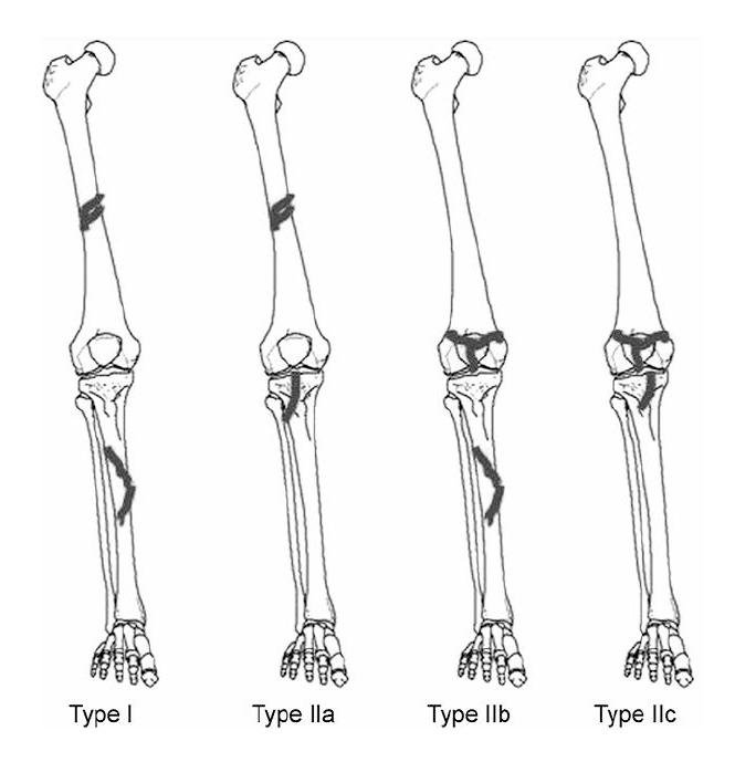
Floating knee injuries may include a combination of diaphyseal, metaphyseal, and intra-articular fractures (see the image below).
[5] This combination of fractures is less common in the pediatric population than in adults. However, epiphyseal injury can adversely affect open growth plates, predisposing a child to limb-length discrepancy and angular deformities.
Classification
Adults
Blake and McBryde used the terms true (or type I) injury and variant (or type II) injury to classify the floating-knee fracture pattern, as follows
[3] :
- Type I is a pure diaphyseal fracture of the femur and tibia
- Type II is a fracture that extends into the knee, hip, or ankle joint [6]
Fraser et al classified floating knee injuries in a similar way by analyzing knee involvement:
-
Type I is the same as the true injury Blake and McBryde described, with extra-articular fractures of both bones
-
Type II is subdivided into three subtypes: type IIa, which involves femoral shaft and tibial plateau fractures; type IIb, which includes fractures of the distal femur and the shaft of the tibia; and type IIc, which indicates fractures of the distal femur and tibial plateau

In both of these classification systems , type II fractures with intra-articular involvement have been linked with higher complication rates and poorer functional results than those observed with type I injuries.
Children
In children, floating knee injuries are classified according to the Bohn-Durbin or Letts classification systems.
In the Bohn-Durbin classification, floating knee injuries are described as follows:
The Bohn-Durbin system does not account for open fractures and cannot be used to predict complications and prognoses.
Unacceptable findings are femoral union in a position of greater than 30° anterior angulation, 15° valgus angulation, and 5° posterior or varus angulation, or greater than 2 cm of shortening. Tibial malunion is defined as greater than 5° angulation in any plane or greater than 1 cm of shortening. Rotational malunion is defined as any internal rotational deformity exceeding findings on the unaffected side or greater than 20° external rotation of the extremity, as detected during walking or standing.
Letts et al designed a classification system in which they recognized diaphyseal, metaphyseal, or epiphyseal knee fractures (types A, B, C) and also open fractures (types D and E) (see the image below). The drawback of this system is that it does not indicate how to classify patients with epiphyseal separation in the distal femur and tibia or how to describe the location of open fractures in the epiphysis, metaphysis, or diaphysis.
[8]
 In both of these classification systems , type II fractures with intra-articular involvement have been linked with higher complication rates and poorer functional results than those observed with type I injuries.
Children
In children, floating knee injuries are classified according to the Bohn-Durbin or Letts classification systems.
In the Bohn-Durbin classification, floating knee injuries are described as follows:
Leave a comment
You must be logged in to post a comment.